Compact Bone Diagram Unlabeled / Bone Structure Human Anatomy And Dissection Reader Openstax Cnx : Its unlabeled, so that your practce better.
byAdmin-
0
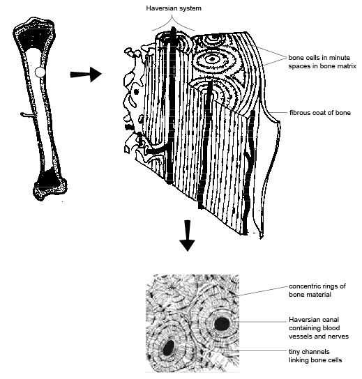
Compact Bone Diagram Unlabeled / Bone Structure Human Anatomy And Dissection Reader Openstax Cnx : Its unlabeled, so that your practce better.. Quizzes on human skeletal system anatomy, bone anatomy, and bone markings. Human gross anatomy study | humandiagram.info. The osteon consists of a central canal called the osteonic (haversian) canal, which is surrounded by concentric rings (lamellae) of matrix. Practice quiz & test prep for students and teachers. Human anatomy physiologyil biol 1611l.
Between the rings of matrix, the bone cells (osteocytes) are located in spaces called lacunae. The bones mentioned in each human skeleton chart are: Skull, clavicle, mandible, scapula, thorax, sternum, humerus, ulna, radius, carpus, phalanges (fingers), metacarpus, spine, pelvis, sacrum, femur, tibia. Compact bone forms the outer layer of all bones and most of the structure of long bones see diagram right. Learn vocabulary, terms and more with flashcards, games and other study tools.
Skull And Vertebral Bone Marrow Are Myeloid Cell Reservoirs For The Meninges And Cns Parenchyma Science from science.sciencemag.org What are diplo , its function, and location? 6 compact bone vs spongy bone. The bones mentioned in each human skeleton chart are: The osteon consists of a central canal called the osteonic (haversian) canal, which is surrounded by concentric rings (lamellae) of matrix. Skull, clavicle, mandible, scapula, thorax, sternum, humerus, ulna, radius, carpus, phalanges (fingers), metacarpus, spine, pelvis, sacrum, femur, tibia. These units allow compact bone to. Printable animal cell diagram u2013 labeled unlabeled and blank. Bone classification, structure & relationships:
Human anatomy physiologyil biol 1611l.
Compact bone consists of outer and inner sheets of lamellar bone (not seen here) and haversian systems, shown here, that run parallel to the long axis of bones. These units allow compact bone to. Key.' carotid canal coronal suture ethmoid bone external occipital protuberance foramen lacerum foramen magnum foramen ovale frontal bone edwnq'p'iep'n glabella. Learn vocabulary, terms and more with flashcards, games and other study tools. Label compact and spongy bone illustrations as demonstrated in class. Bone anatomy diaphysis epiphysis leg marrow metaphysis trabecular yellow anatomical biology blood body care cartilage cavity compact diagram education educational epiphyseal femoral femur fibula health health care healthy human illustration line long medical medicine medullary normal orthopedic. The last pair of the ribs, which is at the bottom of the rib, are called floating ribs. Create your own flashcards or choose from millions created by other students. The bones shown in the chest and hip region in the labeled human skeleton diagram are the ribs, vertebrae, pelvis, os coxae, sacrum and coccyx. Location of red and yellow marrow in adults and. Hand, grasping organ at the end of the forelimb of certain vertebrates that exhibits great mobility and flexibility in the digits and in the whole organ. The bones mentioned in each human skeleton chart are: The outer walls of the diaphysis cortex cortical bone are composed of dense and hard compact bone a form of osseous tissue.
Femur bone diagram unlabeled via. Compact bone, also known as cortical bone, is a denser material used to create much of the hard structure of the skeleton. Start studying fracture repair unlabeled. Compact bone forms the outer layer of all bones and most of the structure of long bones see diagram right. Create your own flashcards or choose from millions created by other students.
Anatomy And Physiology Of Animals The Skeleton Wikibooks Open Books For An Open World from upload.wikimedia.org The long bones of the body contain many distinct regions due to the way in which they develop. This lab is designed to provide students with an overview of bones through a variety of investigative to identify the major regions and structures of an osteon in a histological specimen of compact bone (or diagram or model of one). Human gross anatomy study | humandiagram.info. Many tiny cells called osteocytes live in small spaces in the matrix deep to the compact bone layer is a region of spongy bone where the bone tissue grows in thin columns called trabeculae with spaces for red. Long bone structure diagram and definitions flashcards quizlet. Gallery long bone diagram unlabeled anatomy and physiology. Compact bone diagram osteon compact bone ap pinterest anatomy human anatomy and. It is a bone is one of two kinds of bone tissue that can be found in the compact type of bone wraps around and protects the only other type of bone tissue known as the you should include the histology of compact bone slides with diagram as well into your article.
Human gross anatomy study | humandiagram.info.
Location of red and yellow marrow in adults and. (b) in this micrograph of the osteon, you can clearly see the concentric lamellae and central canals. Hand, grasping organ at the end of the forelimb of certain vertebrates that exhibits great mobility and flexibility in the digits and in the whole organ. Anatomical diagrams for medical students. Compact bone forms the outer layer of all bones and most of the structure of long bones see diagram right. Compact bone, also known as cortical bone, is a denser material used to create much of the hard structure of the skeleton. The outer part of a long bone is made of compact bone. Quizzes on human skeletal system anatomy, bone anatomy, and bone markings. A typical long bone showing gross anatomical features. The bones shown in the chest and hip region in the labeled human skeleton diagram are the ribs, vertebrae, pelvis, os coxae, sacrum and coccyx. The long bones of the body contain many distinct regions due to the way in which they develop. Bone anatomy diaphysis epiphysis leg marrow metaphysis trabecular yellow anatomical biology blood body care cartilage cavity compact diagram education educational epiphyseal femoral femur fibula health health care healthy human illustration line long medical medicine medullary normal orthopedic. Total there are 12 pairs of ribs, as you can see in the diagram.
Edraw is a new uml diagram and software diagram drawing tool. Learn vocabulary, terms and more with flashcards, games and other study tools. As seen in the image compact bone is formed from a number of osteons, which are circular units of bone material and blood vessels. Microscopic bone anatomy human body diagram. Start studying fracture repair unlabeled.
Pin On A P 2 Skin Bone Muscle from i.pinimg.com Femur bone diagram unlabeled via. These units allow compact bone to. Printable animal cell diagram u2013 labeled unlabeled and blank. Unlabeled diagram showing the carpal bones (download free pdf below!) now you've seen the carpal bones labeled and unlabeled, it's time to move on to our interactive carpal bones quizzes. As seen in the image compact bone is formed from a number of osteons, which are circular units of bone material and blood vessels. The outer part of a long bone is made of compact bone. Compact bone consists of outer and inner sheets of lamellar bone (not seen here) and haversian systems, shown here, that run parallel to the long axis of bones. The osteon consists of a central canal called the osteonic (haversian) canal, which is surrounded by concentric rings (lamellae) of matrix.
Location of red and yellow marrow in adults and.
Femur bone diagram unlabeled via. Anchor chart human bone diagram human body skeleton stem science health hand. Bone classification, structure & relationships: Create your own flashcards or choose from millions created by other students. Learn vocabulary, terms and more with flashcards, games and other study tools. Edraw is a new uml diagram and software diagram drawing tool. Long bone structure diagram and definitions flashcards quizlet. A typical long bone showing gross anatomical features. Sclerostin inhibits bone formation mostly by antagonizing lrp5/6, thus inhibiting wnt signaling. Compact bone diagram osteon compact bone ap pinterest anatomy human anatomy and. The outer part of a long bone is made of compact bone. These units allow compact bone to. Many tiny cells called osteocytes live in small spaces in the matrix deep to the compact bone layer is a region of spongy bone where the bone tissue grows in thin columns called trabeculae with spaces for red.
structure of a bone diagram compact bone diagram femur diagram osteon structure of bones what does spongy bone do human anatomy bone function parts of a long bone unlabeled diagram system compact bone diagram. Long bone structure diagram and definitions flashcards quizlet.